
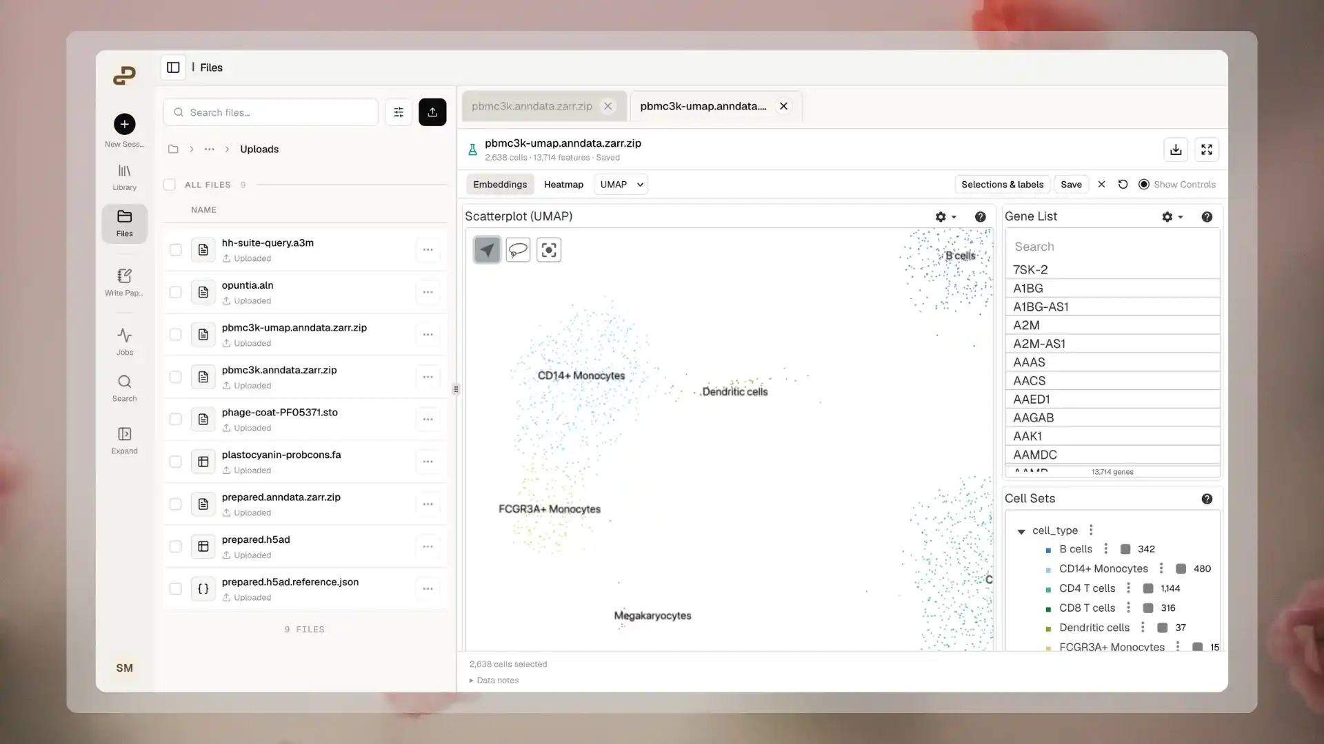
Explore a prepared UMAP embedding alongside gene and cell-group controls.
What you can explore
Only available views are offered. A missing Heatmap or Spatial option can mean that the dataset lacks the required inputs, rather than that loading failed.
Supported files
Extensions are case-insensitive. Keep the correct compound filename, such as
sample.ome.tiff or sample.ome.zarr.zip. Renaming an unrelated file does not convert its contents.
An image-only file provides image viewing, not cell identities, gene expression, or saved cell selections. Uploading an image and an AnnData file separately does not automatically align or link them. Spatial coordinates in an H5AD file alone do not automatically create a linked tissue-image view.
Prepare an AnnData viewing copy
Use an already-processed dataset. Check the following before uploading:- Each cell has a unique, consistent identifier. Embeddings, groups, and expression rows must refer to the same cells in the same order.
- Embedding coordinates are numeric and already computed. Standard UMAP and t-SNE inputs are two-dimensional. For supported PCA data, the viewer displays the first two components.
- Cell-group columns are stored as categorical metadata when you want group controls. A text column is not automatically treated as a cell-type grouping.
- Expression is supplied at the scale you want to inspect. Supported matrices include dense, CSC, and CSR data. For large sparse datasets, a CSC viewing copy can make gene exploration more responsive.
- A heatmap has a prepared boolean feature filter, such as a highly variable gene subset. A full expression matrix does not automatically enable the heatmap.
Package Zarr archives correctly
For AnnData, the ZIP must contain one store at the archive root or inside one top-level folder ending in.zarr.
For OME imagery, place the image store directly at the ZIP root. A wrapper folder is not supported for the direct OME-Zarr image path.
Use ZIP Store mode, also called ZIP_STORED or no ZIP-level compression. Compressing Zarr data internally is different from compressing the surrounding ZIP entries. Do not use a normal ZIP compression setting and assume it will be compatible.
Private multi-file .zarr directories, archives containing multiple stores, and arbitrary collections of files are not direct substitutes for these prepared archives. Other Zarr versions or encodings are not guaranteed to work.
How Purna prepares your file
- Upload completes. Add the file through Files or chat. Wait until the upload finishes.
- Purna checks supported data. For processed H5AD and AnnData-Zarr archives, Purna checks the contents and prepares the information needed to open the available views. For H5AD, upload the dataset itself; you do not need to prepare or upload a companion file.
- The viewer becomes available. While preparation is pending, you may see Preparing viewer…. Open the file when it is ready, or leave its tab open to continue automatically when preparation finishes.
Working with your data
Open and arrange the viewer
- Open a supported file from Files or a chat attachment.
- Wait for preparation and loading to finish.
- Enable Show Controls to reveal available gene, cell-group, or image-layer controls.
- Choose Embeddings, Heatmap, or Spatial when that view is available.
Explore embeddings and expression
- Pan and zoom to inspect a cluster or individual cells.
- Choose another embedding when the dataset contains more than one supported option.
- Search the gene list and select a gene to change expression coloring.
- Expand Cell Sets to explore available groups and their highlighting.
- Open Heatmap to inspect the prepared gene subset when available.
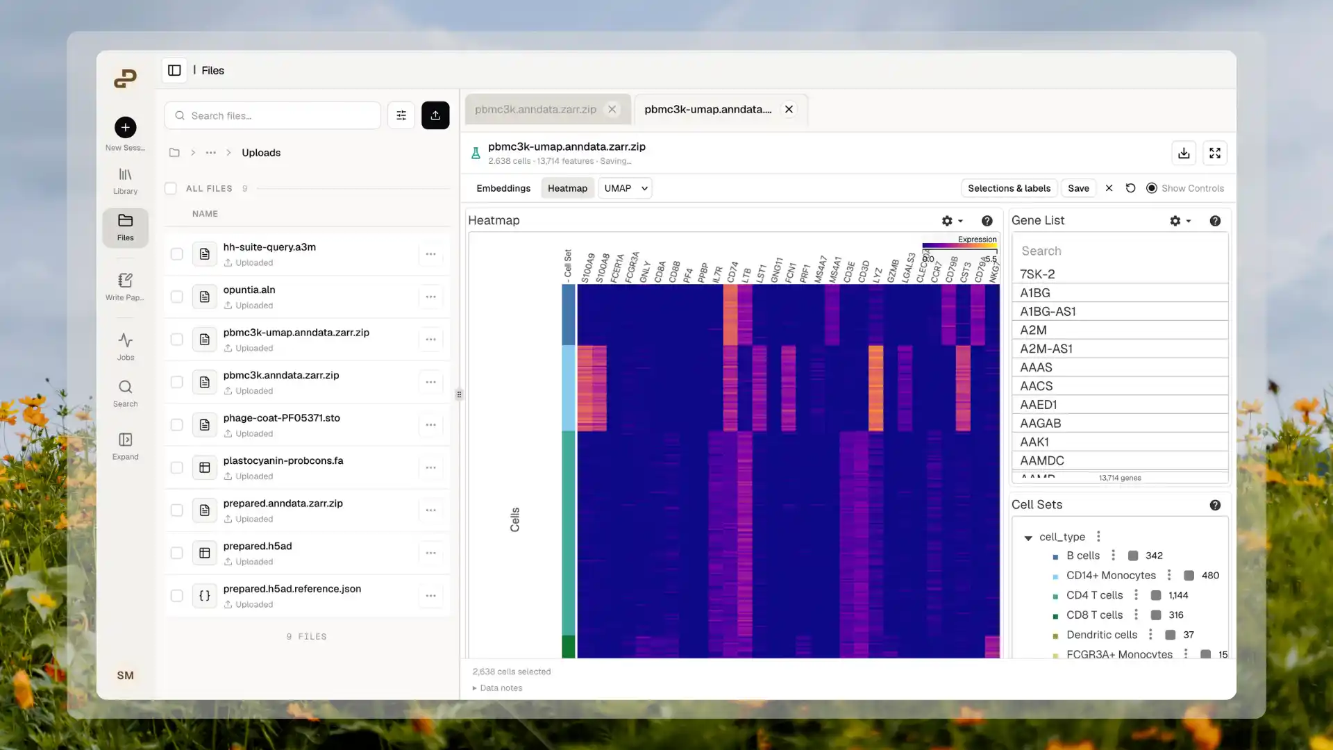
Switch to Heatmap to inspect expression across the prepared gene subset.
Select cells or a region
Use the plot’s lasso tool to draw around cells, or select an existing group in Cell Sets. Wait for cell identities to load, then check the selected-cell count. Use Clear selection to start again. Where a configured spatial view provides cell-selection tools, you can select cells in a spatial region and compare highlighting across its linked views. This requires prepared cell identities and matching spatial coordinates; an image alone cannot provide that information. A saved cell selection records the cells you selected. It is not a saved tissue-boundary drawing, freehand image annotation, or segmentation edit. The current viewer does not provide a general image-painting or region-boundary editor.Save selections, labels, and notes
For a prepared dataset saved in Files with cell-selection controls:- Select the cells you want to keep.
- Open Selections. Some versions label this panel Selections & labels.
- Choose Save selection and give the selection a meaningful name, such as
Outliers to review. - Open that selection’s details to edit its cell label or note. In the compact panel, use the pencil icon; Done closes the editor.
- Wait until the viewer reports Saved before closing the tab or reloading.
What is saved
For supported prepared files opened from Files, personal viewer settings can include the selected embedding or gene, camera and color settings, layout, and controls visibility. Where cell-selection controls are available, named selections, cell labels, and notes also persist after closing and reopening the file or reloading the page, once the save succeeds. These updates belong to you and the particular version of the source file. They are not collaborative edits to the original dataset. Replacing the source starts a new view; Purna does not silently assign old labels to a replacement file. Downloading the original does not include your personal overlays. Local configurations, custom configuration-file sessions, and demonstrations remain temporary. Do not assume their interactions will return after closing or reloading.Work with an agent
Agents can work with your files to help with analysis, statistics, and scripts. They can read the cell identities, labels, and notes in a selection you explicitly share. They can also help write plotting scripts, but you must inspect the resulting graphics yourself. To share a selection, open the dataset in a saved chat in the same project and choose Attach to chat or Attach selection to chat, depending on your version. This adds the selection and source file to the composer. Add your question and send it yourself; attaching does not automatically send a message or start an analysis. The shared file contains the selected cells and overlapping labels and notes. Treat this as sharing those details with the project. Your other personal selections are not included automatically. Select the cells you intend to discuss and attach a new snapshot after changing them. For example, you can ask:- “Read the attached selection and source dataset. Report how many selected cells belong to each supplied cell group.”
- “Write a reproducible script comparing expression in these selected cells with the remaining cells. State the normalization, sample grouping, and statistical assumptions before interpreting the result.”
- “Check whether these saved cell identifiers still match the source file before continuing.”
Limits and unsupported inputs
Preview limits are separate from upload limits. A successful upload does not guarantee comfortable viewing of a large dataset. Large cell counts, dense expression, image channels, or complex metadata can take longer to open and consume more browser memory. Use a smaller prepared viewing copy if the browser becomes unresponsive. A missing or oversized heatmap feature subset can leave the heatmap unavailable while other views still work.
The current viewer does not directly open or automatically prepare:
- Standalone CSV, TSV, or spreadsheet data as an omics view. CSV keeps its table preview; existing embedding coordinates must be packaged into a compatible AnnData viewing file.
- Raw counts with neither a supported embedding nor a usable prepared heatmap. Preparation does not calculate the missing analysis.
- General HDF5
.h5files, raw sequencing reads, standalone MTX or raw 10x bundles, Seurat.rdsobjects, or.loomfiles as native omics inputs. - Arbitrary ZIP or TAR archives, multiple-store ZIPs, ZIP entries using unsupported compression, incomplete archives, or renamed files whose contents do not match their extension.
- Private multi-file Zarr folders as direct upload-and-open datasets.
- Ordinary TIFF or general DICOM slides through this viewer. Supported SVS and NDPI files open in the Slides Viewer. A standard
.tifis not an OME-TIFF simply because it has been renamed. - H5AD files that rely on external data files or unsupported encodings, or inputs that exceed preparation limits.
- Custom viewer plugins, arbitrary configurations, or the full set of controls available in other specialized scientific applications.
Troubleshooting
Return to Scientific Components, or see Files for uploading and organizing your data.